Upper Leg Tendon Anatomy - Quadriceps Thighs Upper Leg A Step Beyond Massage Therapy - Spicermanyt at checkout for 40% off this tutorial!
byAdmin•
0
Upper Leg Tendon Anatomy - Quadriceps Thighs Upper Leg A Step Beyond Massage Therapy - Spicermanyt at checkout for 40% off this tutorial!. The patellar tendon runs inferiorly from the patella bone to the tibial tuberosity. This may result in tendon subluxation; They are remarkably strong, having one of the highest tensile strengths found among soft tissues. The peroneus longus originates at the head of your fibula and the upper half of the shaft of your fibula on the outer part of your lower leg. When a muscle contracts, the tendon pulls on the bone causing the joint to move.
Tendons are fibrous cords attached to muscles and bone. Concept 3d illustration back upper leg human anatomy. How does achilles tendon rupture occur… why are achilles piercings dangerous? Lie prone on a hamstring curl machine. The tendons of the edl can be palpated on the dorsal surface of the foot.
Conceptual 3d Human Front Upper Leg Muscle Anatomy Buy This Stock Illustration And Explore Similar Illustrations At Adobe Stock Adobe Stock from as1.ftcdn.net Tendons transmit the mechanical force of muscle contraction to the bones. 3d illustration back fit strong human anatomy. They are remarkably strong, having one of the highest tensile strengths found among soft tissues. Lie prone on a hamstring curl machine. By spicer mcleroy in tutorials. Injuries to the achilles tendon are very serious. Lateral (fibular) collateral ligament (fcl) upper part middle part lower part popliteus tendon (pt) upper part i. An anatomical and biomechanical study.
Tendons are fibrous cords attached to muscles and bone.
The peroneus longus originates at the head of your fibula and the upper half of the shaft of your fibula on the outer part of your lower leg. When a muscle contracts, the tendon pulls on the bone causing the joint to move. Spicermanyt at checkout for 40% off this tutorial! There are four muscles in the anterior compartment of the leg. The tendons of the edl can be palpated on the dorsal surface of the foot. Concept 3d illustration back upper leg human anatomy. And it is also critical to the walking process. Lie prone on a hamstring curl machine. Use the mouse scroll wheel to move the images up and down alternatively use the tiny arrows (>>) on both side of the image to move the images. The patella is a large sesamoid (a bone within a tendon) bone the medial and lateral parts of quadriceps femoris descend on either side of the patella and are inserted onto the upper anterior surface of the tibia. Tendons are fibrous cords attached to muscles and bone. It is the largest tendon of the parts of leg. The human leg, in the general word sense, is the entire lower limb of the human body, including the foot, thigh and even the hip or gluteal region.
Tendon, tissue that attaches a muscle to other body parts, usually bones. There is no real division between the core and the upper leg; By spicer mcleroy in tutorials. Palmar region , arteries (illustrations: 630 anatomical structures of the upper limb (pectoral girdle, shoulder, arm, elbow, forearm, wrist, hand and fingers) were labeled.
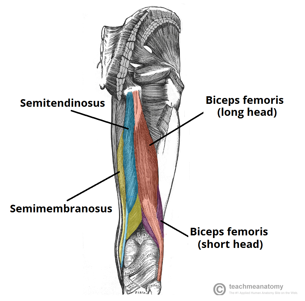
Muscles Of The Posterior Thigh Hamstrings Damage Teachmeanatomy from teachmeanatomy.info The patella is a large sesamoid (a bone within a tendon) bone the medial and lateral parts of quadriceps femoris descend on either side of the patella and are inserted onto the upper anterior surface of the tibia. Tendon, tissue that attaches a muscle to other body parts, usually bones. Lateral (fibular) collateral ligament (fcl) upper part middle part lower part popliteus tendon (pt) upper part i. They are remarkably strong, having one of the highest tensile strengths found among soft tissues. Superficial veins of upper limb , anatomy : In this upper leg tutorial, i go over all the major points of the upper leg to take your sculpting skills. Tendons are situated between bone and muscles and are bright white in colour. Use the mouse scroll wheel to move the images up and down alternatively use the tiny arrows (>>) on both side of the image to move the images.
Spicermanyt at checkout for 40% off this tutorial!
Originates from the lateral condyle of the tibia and the medial surface of the fibula. When a muscle contracts, the tendon pulls on the bone causing the joint to move. Palmar region , arteries (illustrations: Collectively, they act to dorsiflex and invert the foot at the ankle joint. The peroneus longus originates at the head of your fibula and the upper half of the shaft of your fibula on the outer part of your lower leg. Related posts of muscle anatomy upper leg. Leg muscle anatomy chart | amulette. Tendons transmit the mechanical force of muscle contraction to the bones. This may result in tendon subluxation; There are four muscles in the anterior compartment of the leg. It is the largest tendon of the parts of leg. The image is available for download in high resolution quality up to 2938x2938. It is formed when the soleus muscle tendon joins with the gastrocnemius tendon.
Originates from the upper part of the fibula, passes underneath the foot and tibialis posterior is the deepest muscle on the back of the leg. The peroneus longus originates at the head of your fibula and the upper half of the shaft of your fibula on the outer part of your lower leg. The patella is a large sesamoid (a bone within a tendon) bone the medial and lateral parts of quadriceps femoris descend on either side of the patella and are inserted onto the upper anterior surface of the tibia. Suspensory ligament of the axilla. Spicermanyt at checkout for 40% off this tutorial!
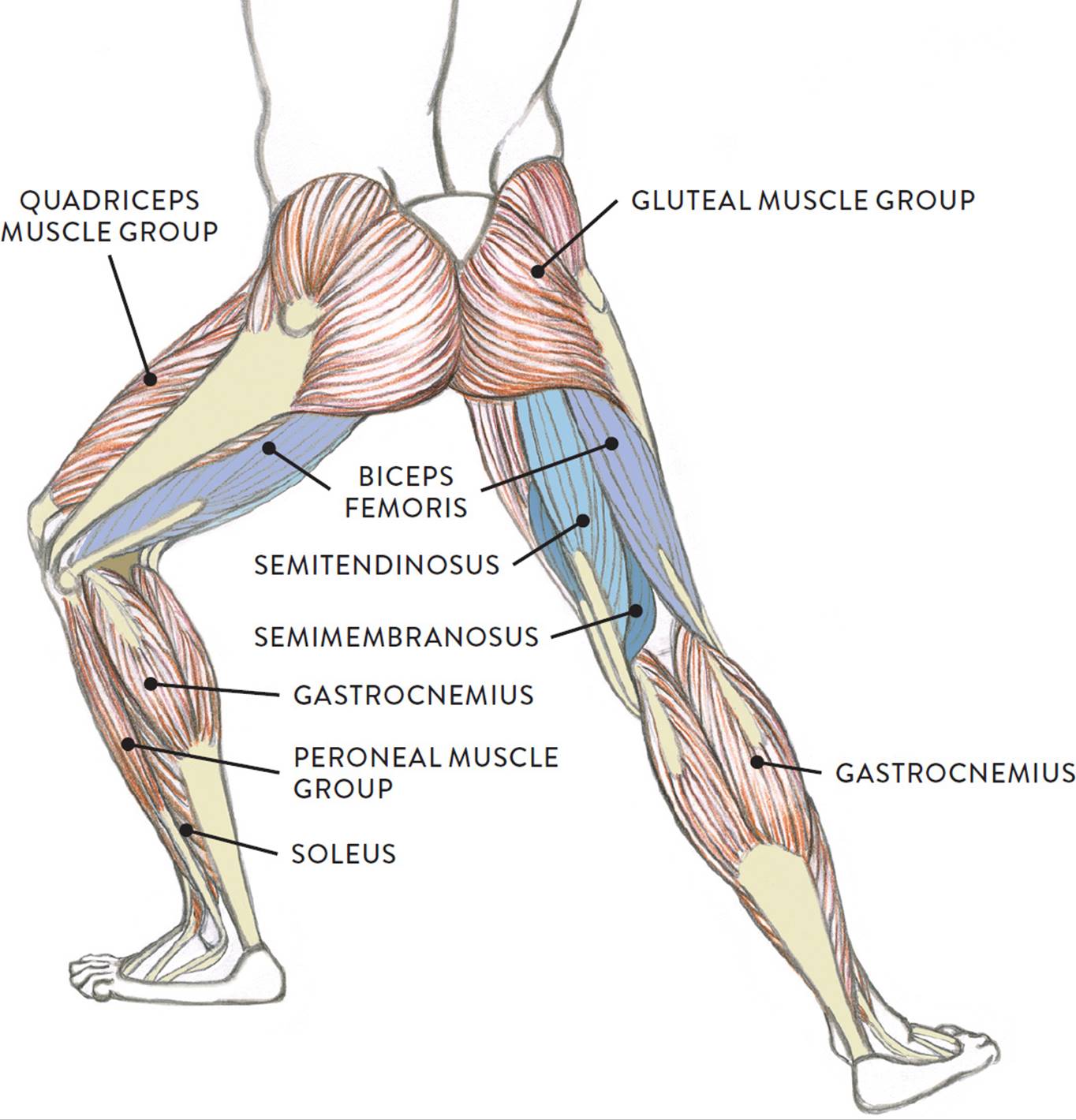
Muscles Of The Leg And Foot Classic Human Anatomy In Motion The Artist S Guide To The Dynamics Of Figure Drawing from doctorlib.info It is formed when the soleus muscle tendon joins with the gastrocnemius tendon. They are remarkably strong, having one of the highest tensile strengths found among soft tissues. Injuries to the achilles tendon are very serious. 3d illustration back fit strong human anatomy. The human leg, in the general word sense, is the entire lower limb of the human body, including the foot, thigh and even the hip or gluteal region. Leg anatomy muscles and tendons how to fix achilles. An anatomical and biomechanical study. Superficial veins of upper limb , anatomy :
Tendons are fibrous cords attached to muscles and bone.
Tendons are thick bands of tissue that connect muscles to bone. 3d illustration back fit strong human anatomy. Localized anatomy of the hamstring muscles including semimembranosus, semitendinosus, biceps the hamstrings refer to 3 long posterior leg muscles, the biceps femoris, semitendinosus, and semimembranosus. There is no real division between the core and the upper leg; Fascia of the upper limb. 1280 x 1520 jpeg 166 кб. .16 penile numbness and perineum tenderness.18 any suggested exercises or stretches?.22 leg musculature 209 elbow tendonitis and saddle sores. This mri wrist coronal cross sectional anatomy tool is absolutely free to use. Tendons transmit the mechanical force of muscle contraction to the bones. Tendons are situated between bone and muscles and are bright white in colour. Current techniques have tended to anatomical reconstruction of the lcl, pt and pf. Related posts of muscle anatomy upper leg. Lie prone on a hamstring curl machine.